We are - world-class medicine more accessible than in Europe and Central Asia
03
Doctors with international
practice
Transfer to and
from Tyumen
Permanent support of the personal
coordinating therapis
Full diagnostics
of the organism
Tourist programme
of Tyumen historical places.
Pleasant stay
of citizens of other countries
How to contact us?
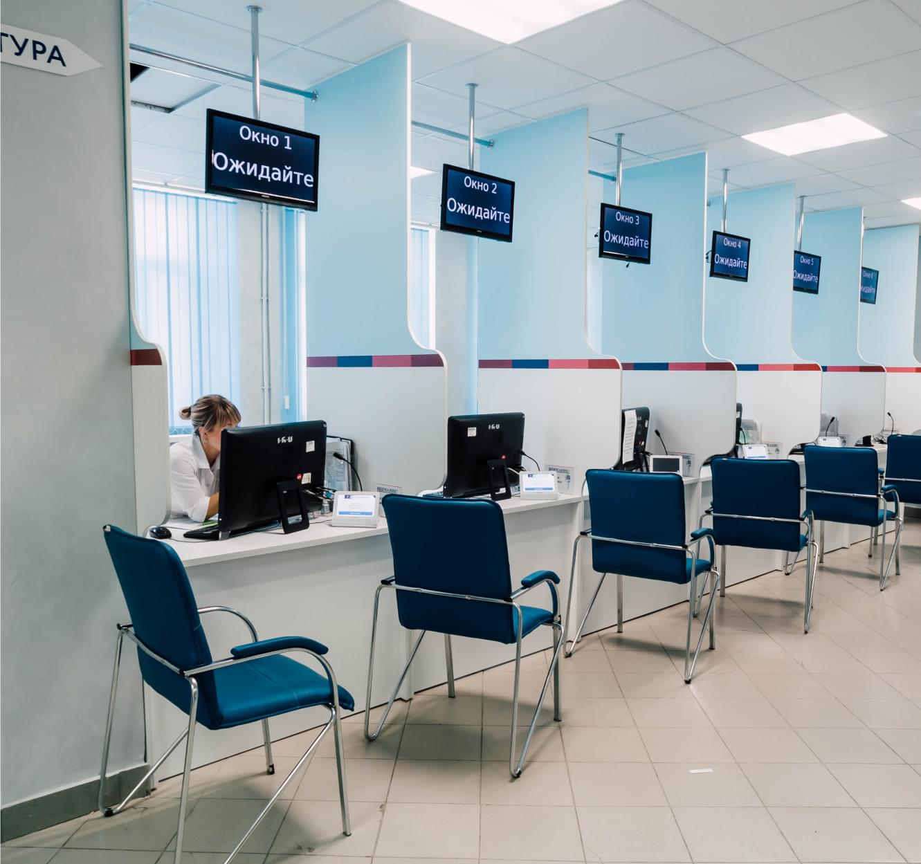
We have established the Medical Tourism Development Coordinating Centre in order for tourists to receive medical services conveniently. The goal of the “MEDICAL CITY” Centre is to make your treatment and stay in Tyumen as comfortable as possible to achieve the result that will exceed all your expectations.
Send medical documentation and MEDICAL CITY's specialists will offer you an individual solution
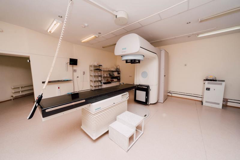
NUCLEAR (radionuclide) DIAGNOSTICS
Radionuclide diagnostics is one of the modern methods of radiation diagnostics for assessing the functional state various organs and body systems using diagnostic radiopharmaceuticals labeled with radionuclides.
Radiopharmaceuticals are certain chemical or biochemical compounds labeled gamma-emitting radionuclides having a short half-life. Gamma radiation coming from the patient’s body, it is registered by the gamma camera detector and after computer processing the received information is converted into functional image of the investigated organ. Spatio-temporal picture of the distribution of the radiopharmaceutical gives an idea of the shape, size and position of the organ, as well as the presence of pathological foci in it.
Radionuclide diagnostics are widely used in oncology, endocrinology, cardiology, urology, neurology, traumatology.
NUCLEAR DIAGNOSTIC METHODS:
1. Scintigraphy of various organs and systems - a method of radionuclide examination of internal organs, based on visualization using a scintillation gamma camera distribution distributed into the body radiopharmaceutical drug.
With static scintigraphy, a two-dimensional image is obtained when one or more scintigrams are performed for studying the anatomical and topographic state of internal organs and detecting foci of pathological distribution of the radiopharmaceutical. This method is most often used for diseases of the thyroid and parathyroid glands, kidneys, liver, lungs.
In dynamic scintigraphy, a series of two-dimensional images is obtained by registering individual frames with given time interval, which allows to determine the nature of the movement of the radiopharmaceutical in the organ under investigation and evaluate its function. The methods of dynamic scintigraphy include research urinary kidney function (dynamic renoscintigraphy), biliary function of the liver and bile bladder (dynamic hepatocholecystography).
2. Whole body scintigraphy in the “Whole body” mode - obtaining an image of the whole body using specialized gamma camera with a large field of view. The advantage of this method is to obtain whole body scintigrams in one study after a single administration of a radiopharmaceutical. Most often used in oncology to identify the primary tumor focus and search for distant metastases, planning and evaluation of treatment results.
3. Single-photon emission computed tomography (SPECT) - allows you to get a layered picture the distribution of the radiopharmaceutical in the organ with the subsequent reconstruction of its three-dimensional image. With new image acquisition technology involves one of the most interesting aspects of quantitative SPECT - the ability to calculating the volume of functioning organ tissue by summing the volumetric elements that form the images organ slices. This modern method is best used in oncology and cardiology.
4. Single-photon emission computed tomography combined with x-ray computed tomography (SPECT / CT) is the latest method of integrated radiation-radiological research, allowing you to simultaneously see not only the inclusion of the radiopharmaceutical in any pathological process, organ, especially with cancer, but also accurately determine the spatial localization of the picture tomographic slice, which significantly improves the quality of scintigraphic images and improves accuracy diagnostics. Such studies are carried out on a modern combined apparatus that combines single-photon emission tomograph and X-ray computer tomograph. This modern technology is perfect. Suitable for tasks related to imaging tumors and planning therapeutic procedures, as well as for examination of cardiological patients.
5. Positron emission computed tomography (PET / CT) is able to diagnose those tumor foci, which cannot be detected using other imaging tools - ultrasound, CT, MRI, radiography. Received in the result of the examination, the results will help to identify neoplasms at the earliest stages, choose an effective treatment tactics.
PET / CT with glucose (18 FDG) - scanning of the whole body when it is necessary to assess the condition of organs and systems. The purpose of the study is to detect pathological processes in the body. Most often performed with the need to identify: a primary tumor, metastases, evaluation of the effectiveness of treatment, relapse of the disease.
PET / CT with 11C-meteonin is used to diagnose malignant neoplasms of the brain, evaluating the effectiveness of treatment, as well as for differential diagnosis between benign and malignant neoplasms of the brain.
PET / CT with 11C-choline is used to evaluate the effectiveness of treatment for prostate cancer, to identify parathyroid adenomas
Subscribe
Stay updated on our news
Receive news about discounts and promotions by email
Телефон
Сайт
Get a consultation
Send medical documentation and MEDICAL CITY's specialists will offer you an individual solution
Заявка не отправлена
Произошла ошибка на сервере
Ваша заявка была отправлена
Наши специалисты свяжутся с вами в ближайшее время